If fear has kept you from taking charge of your dental problems, you have now taken the bravest step: addressing the problem and looking for the solution.
We know how difficult this decision is, which is why dental problems are treated in our clinic with the greatest possible empathy and care. Of course, this also applies to oral surgery treatments.
Oral surgery is an independent and very complex area of dentistry, therefore only experienced and highly qualified specialists carry out these procedures in our clinic.
Complicated tooth removals
There are cases where tooth removal is a complex task and can only be carried out through surgery.
This can happen, for example, if the tooth is covered by gum or bone, but it also often happens that the structure of the tooth is severely damaged, for example parts of the tooth are already missing, or it broke due to extensive caries, an injury or an accident.
In these cases, the tooth and tooth remains are removed surgically.
Wisdom tooth removal
A very common oral surgical procedure is the removal of wisdom teeth, especially in cases where the tooth cannot break through the gums without the appropriate application of dental surgery.
Similar to the other molar teeth, the purpose of the wisdom tooth is to chew food and grind it properly. Wisdom teeth have a special structure: in some cases they are single-rooted, in other cases they can be multi-rooted, and it is not uncommon for a wisdom tooth to even have five roots.
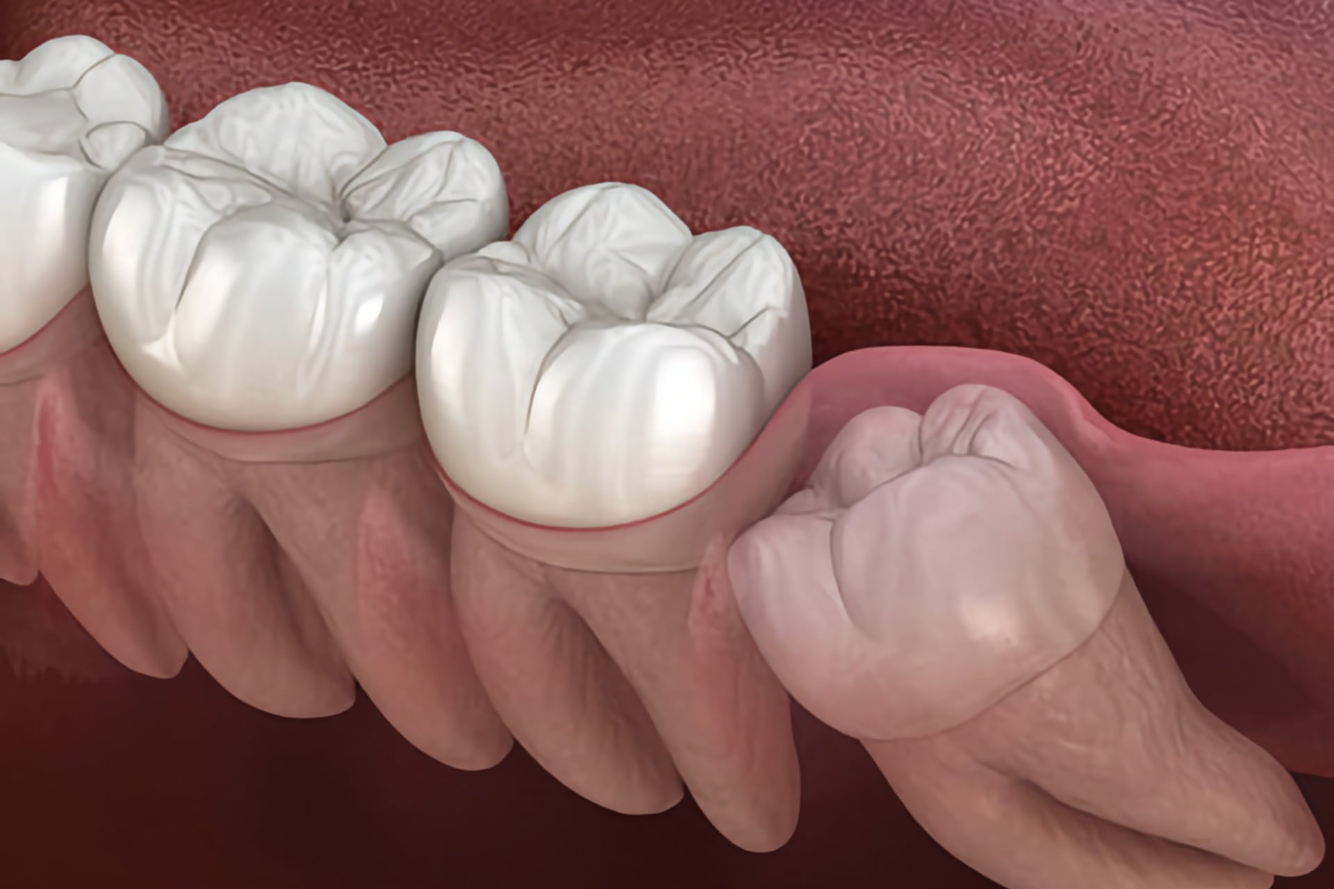
It is a common evolutionary problem that wisdom teeth cannot grow out of the gums, which in many cases is not a problem, but surgical removal of wisdom teeth is still necessary if, for example, the stuck wisdom teeth push the other teeth inwards and therefore the teeth are overloaded, which can lead to the teeth shifting.
It can also happen that a wisdom tooth needs to be extracted even though it has erupted through the gums normally. Its location makes it difficult to clean, so tartar easily sticks to its surface and bacteria can grow on it.
The removal of wisdom teeth is considered a routine procedure. It can be carried out completely painlessly under local or general anesthesia.
Oral surgical removal of the root tip – root tip resection
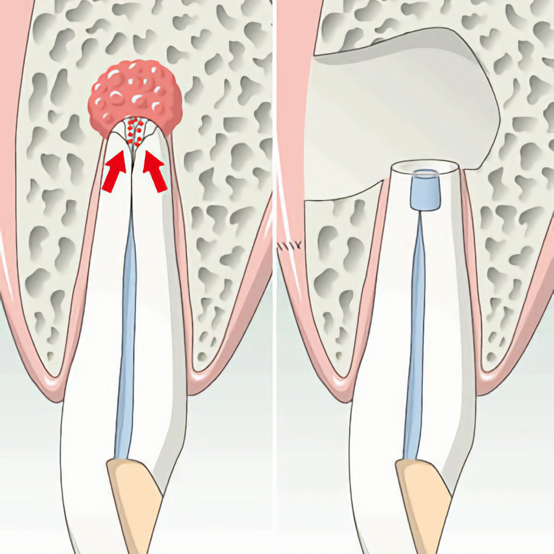
If a cyst or another disorder is found at the root tip in the bone, in most cases it is a sign that the dental nerve in that tooth has died off. This condition does not cause any discomfort for a long time, so people do not notice this development, although in some cases the problem draws attention to itself through tooth discoloration or sensitivity when biting.
If root canal treatment is unsuccessful or not possible for any reason and the inflamed tissue needs to be removed, a surgical removal of the root tip is necessary. If the infected root tip is not removed in time, it can spread and lead to tooth loss, which is why a quick oral surgery is important.
Bone graft
Missing teeth that have not been replaced for a long period of time lead to severe bone loss from year to year, affecting the stability and strength of the jawbone.
The options for dental implantation are determined by the patient’s bone structure: The prerequisite is sufficient bone height and bone width so that an implant can be safely inserted and stably and permanently integrated into the jawbone. Here too, the trend is towards minimally invasive procedures that cause little pain and ensure a fast recovery.
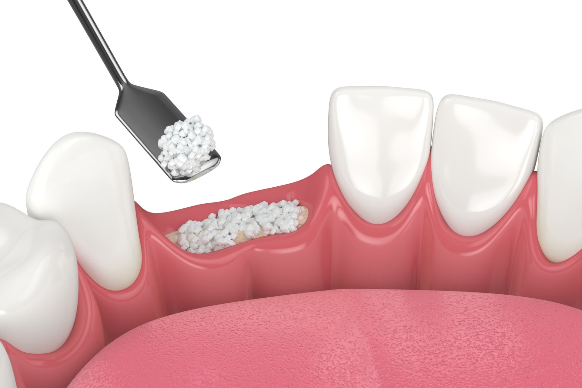
A detailed preliminary examination is always carried out before implantation. The amount of bone present in the mouth can only be assessed as part of a personal preliminary examination – supported by a panoramic X-ray and/or a 3D CT image. Sufficient bone material for long-term reliable implant solutions starts with a bone width of at least 5 mm and a bone height of at least 9 mm.
However, the need for bone grafting may be justified even if sufficient bone is present. Although CT diagnostics represent the bones in three dimensions and thus enable precise measurements, the implantologist only learns some aspects about the quality of the bone during the procedure.
In unfavorable conditions (e.g. porous bone), bone augmentation may be necessary so that the implant is firmly anchored in the jawbone.
The bone structure enables different interventions on both jawbones, the lower and upper jaw, and thus enables tooth replacement with implants even for those patients for whom a fixed solution on implants would otherwise not be possible.
Sinus lift
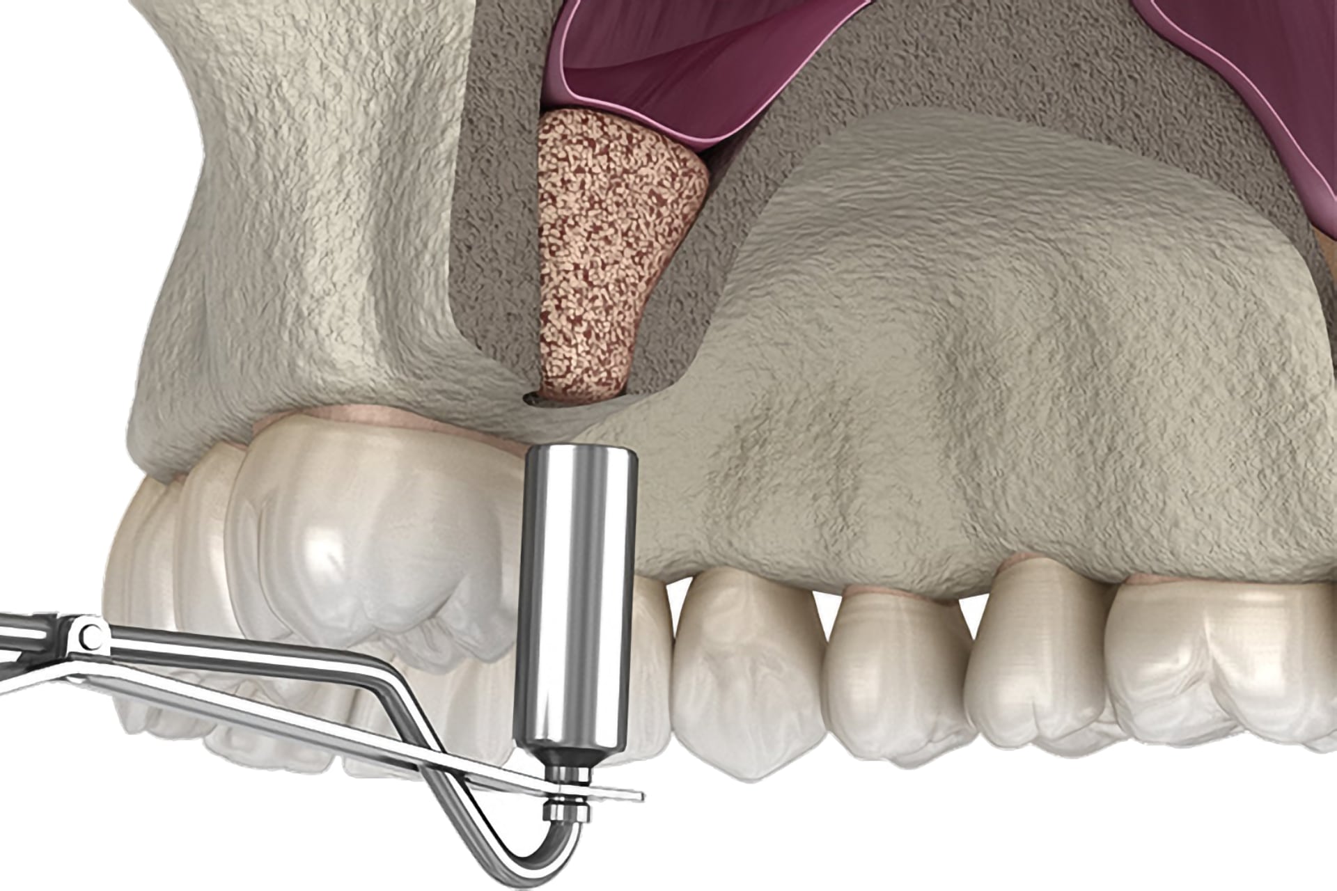
The sinus lift is a popular and frequently used surgical technique when there is not enough bone material of sufficient size for dental implants in the back part.
With its help, artificial bone can be built up under the facial cavity, stimulating the natural bone to grow again while dissolving, resulting in a high chance that a dental implant can be inserted.
The sinus lies above the area of the upper chewing teeth.
The molars are the first teeth to grow in childhood, so in adulthood these teeth have already decayed in many cases or have been filled several times, so these are usually the first to be lost or coming into the need of being removed.
If a lot of time passes after tooth extraction or tooth loss, the bone at the site of the removed tooth begins to disappear, which also means that the lower edge of the maxillary sinus approaches the upper edge of the gums.
This position is extremely important in implant treatment, as implants in the position of the molars are fundamentally important for stabilization. Because primary stability must be achieved in this section, dental implants must be screwed in relatively deeply in the molar area so that the implant-carried tooth can withstand the strong chewing force for a long time. Ideally an implant screw with at least 2 cm length and 4.5-5 mm width should be used in the molar area.
However, the reality is that after the first molars are removed, the remaining bone size is usually only a fraction of that, so the bone needs to be thickened as described above.
During the sinus lift process, an area of the facial cavity is filled with bone substitute material in order to gain the desired bone mass.
PRF method
Platelet-rich fibrin (PRF) is a natural substance found in the blood.
The main point of the procedure is that tissue is obtained from the patient’s own blood as a replacement material to replace the tissue-deficient area created during oral surgery procedures (tooth extraction, cyst removal, etc.) or to regenerate receding gums due to periodontitis. For this purpose the regeneration cells and factors in the blood are used in a very efficient way.
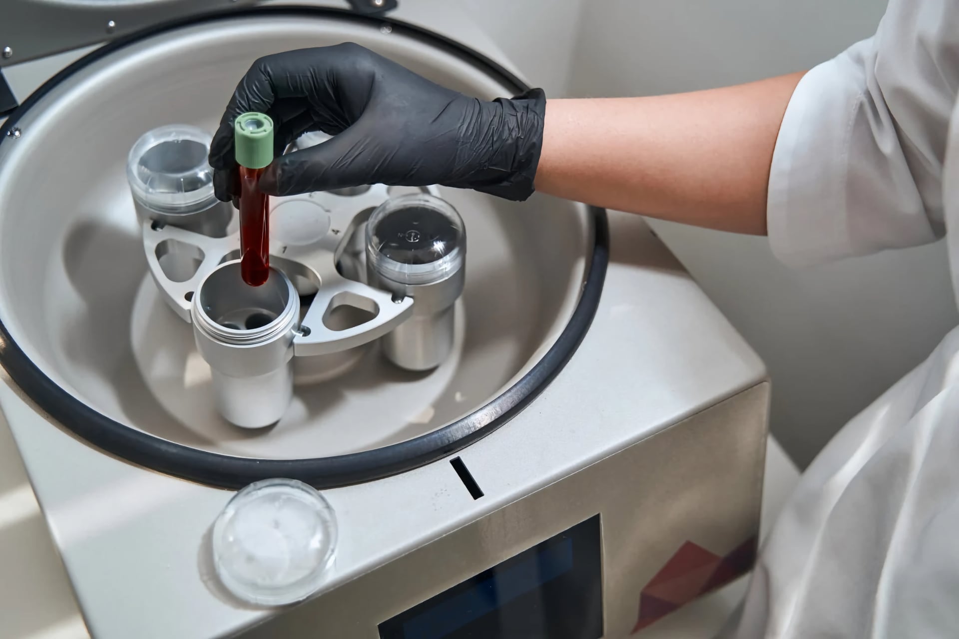
For patients who are waiting for a dental implantation but do not have sufficient bone stock, the PRF method can be used to obtain a substance suitable for bone replacement as an auxiliary material. This is then used alone or – in the case of severe bone deficiency – mixed with artificially produced bone replacement material.
Its huge advantage is it’s 100% efficiency since it is extracted from the patient’s own body.
The procedure to apply PRF is simple and quick.
Advantages of the PRF
- Completely biocompatible.
- Easy to use, low cost.
- Quick production: approx. 20 minutes.
- No risks for the patient as only their own blood is required and used for the process.
- Accelerated wound healing and accelerated tissue development.
- Better wound healing in soft tissue and bones.
- Reduction of the risk of infection after tooth extractions.
